
Imaging is how we stop guessing. Digital radiography and ultrasound let us see inside your pet without surgery, in-house and usually within the same visit — which matters most on the days when waiting for an answer is the hardest part.
What we can see, and when we use it
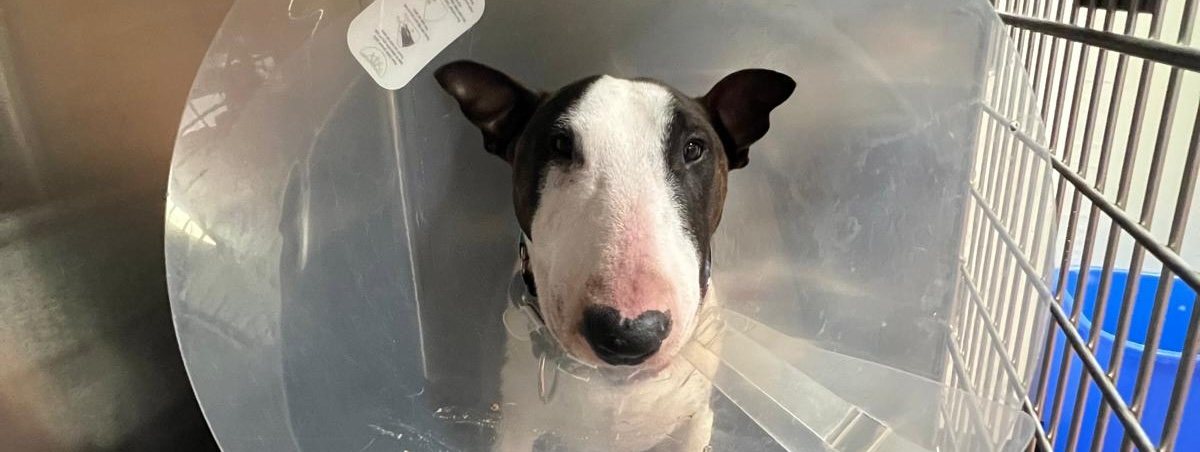
Radiographs are excellent for bone, chest, and the overall shape and position of organs — fractures, arthritis, heart size, foreign bodies, bladder stones. Ultrasound shows soft tissue architecture in a way x-rays cannot: the internal structure of the liver, kidneys, spleen and intestinal wall, and movement in real time.
They answer different questions, and often we want both. What we will not do is run every test available because it exists. Not every test is necessary, and we will tell you which ones will actually change what we do next.
- Digital radiography for timely, high-quality results
- Ultrasound for pregnancy diagnosis and abdominal evaluation
- OFA, PennHIP, and SV/WUSV international x-ray submissions
- Thyroid, basic cardiac, patella and dentition clearances
- Shoulder, spine, and trachea radiographs
- In-house results for immediate clinical decisions
Health clearances and breeder radiographs
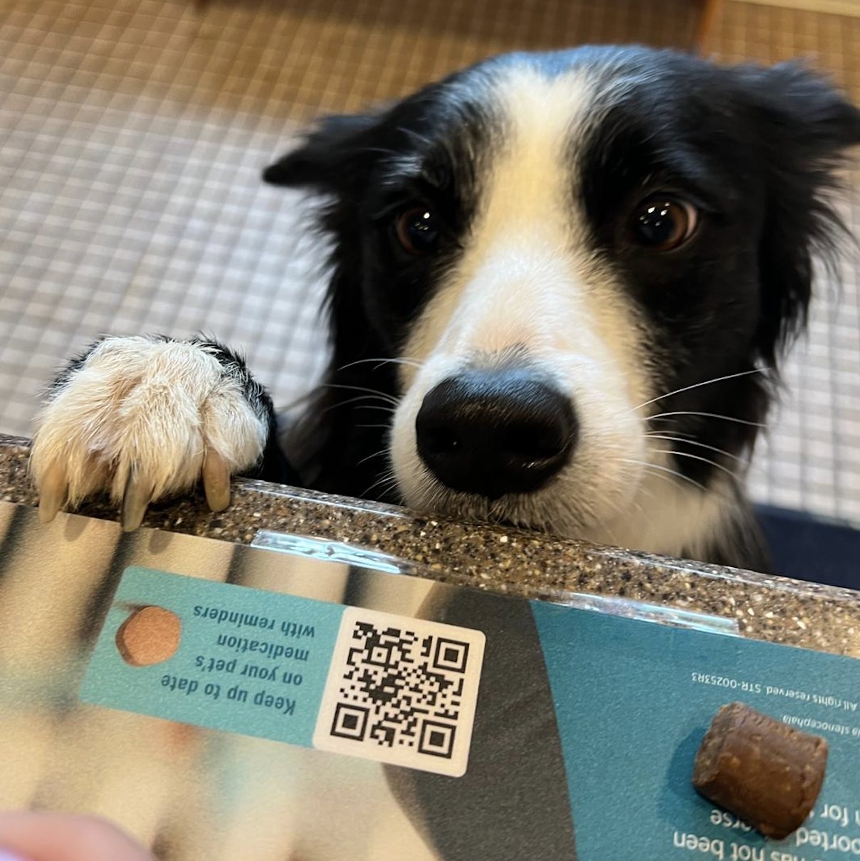
We take OFA hip and elbow radiographs regularly, and those can often be positioned without sedation — which breeders tell us is unusual and appreciated. Correct positioning is everything: a poorly positioned hip can produce a grade that does not reflect the animal, with real consequences for a breeding program.
We also perform PennHIP radiographs, which measure joint laxity and can be done considerably earlier than an OFA evaluation, and we prepare SV/WUSV international submissions for German Shepherd breeders working to the German standard. Both of those schemes require sedation, because each depends on positioning that cannot be achieved safely or accurately in an awake dog. Between the three, we can support whichever scheme your registry or your breed club requires.
Beyond hips and elbows we also handle thyroid, basic cardiac, patella and dentition clearances, along with shoulder, spine, and trachea radiographs. The only clearances we do not offer are those requiring a specialist — eye certification and advanced cardiac.
It is patient work, and we would rather take the extra time than send back a submission that does the dog a disservice.
Answers while you are still here
Because imaging and laboratory work are done in-house, we can often move from concern to diagnosis to plan within a single appointment. That is partly convenience and mostly medicine — an obstruction, a bleeding abdomen, or a pregnancy in trouble are all situations where hours matter.
Common questions
Will my pet need sedation for x-rays?
For most everyday radiographs, often not — many patients hold still enough with gentle handling, and OFA hip and elbow films can frequently be taken without it. PennHIP and SV/WUSV submissions are the exception and do require sedation, since both depend on positioning that cannot be achieved safely in an awake dog. Sedation is also used when positioning would otherwise be painful or when a patient is too anxious to be still safely.
What is the difference between an x-ray and an ultrasound?
Radiographs show bone and the size, shape, and position of organs, making them ideal for fractures, chest evaluation, and foreign bodies. Ultrasound shows the internal structure of soft tissue and movement in real time, making it better for evaluating organ architecture, pregnancy, and fluid. They answer different questions and are frequently used together.
Are x-rays safe for pets?
Yes. The radiation dose from diagnostic radiographs is very low, and digital systems require less exposure than older film. Staff follow strict safety protocols including protective equipment, and the diagnostic benefit substantially outweighs the minimal risk of occasional imaging.
How soon will we have results?
Because imaging is performed in-house, radiographs and ultrasound are typically reviewed during your visit, so you usually leave with findings and a plan rather than waiting for an outside report. Complex studies are sometimes sent for specialist interpretation, which takes longer.
When can pregnancy be detected by ultrasound?
Ultrasound can generally confirm pregnancy in dogs from around 25 to 30 days after breeding and can assess fetal heartbeats and viability. Radiographs are better for counting puppies accurately, but only later in gestation once fetal skeletons have mineralized, typically after about day 45.
References & further reading
The guidance on this page follows current veterinary consensus. These are the sources behind it, if you would like to read further.
- Hip and elbow radiograph requirements — OFA
- Diagnostic imaging in veterinary practice — Merck Veterinary Manual
- American College of Veterinary Radiology — ACVR